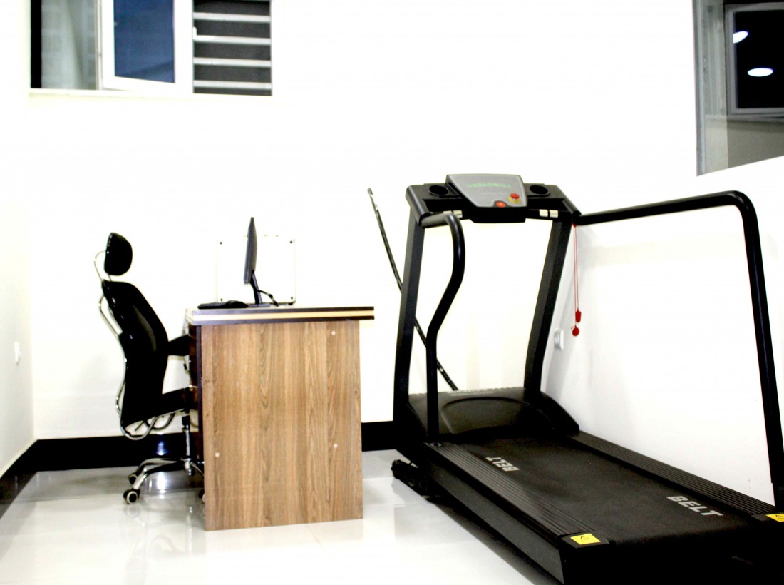
COVID-19 Laboratory
Zwan Medical Complex's Covid-19 PCR test accepted across all the flight agencies in Afghanistan. What is a PCR test? PCR… ...
Read MoreContact Us For Help
4 , sherpur street 7 at the right side from sherpur square, kabul, afghanistan.
Covid 19 Testing Center
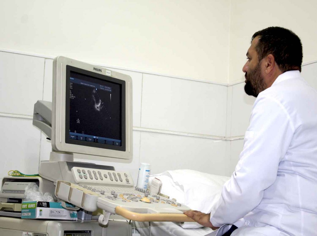
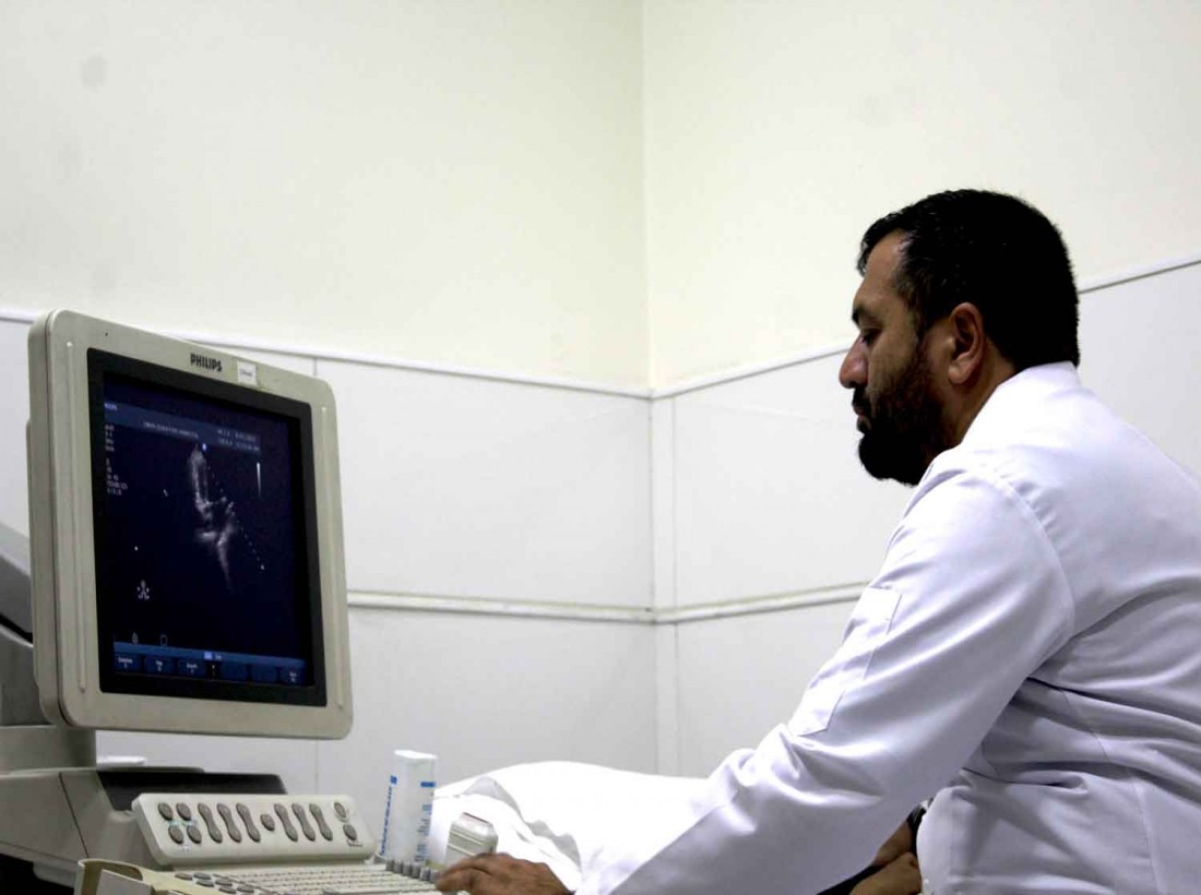
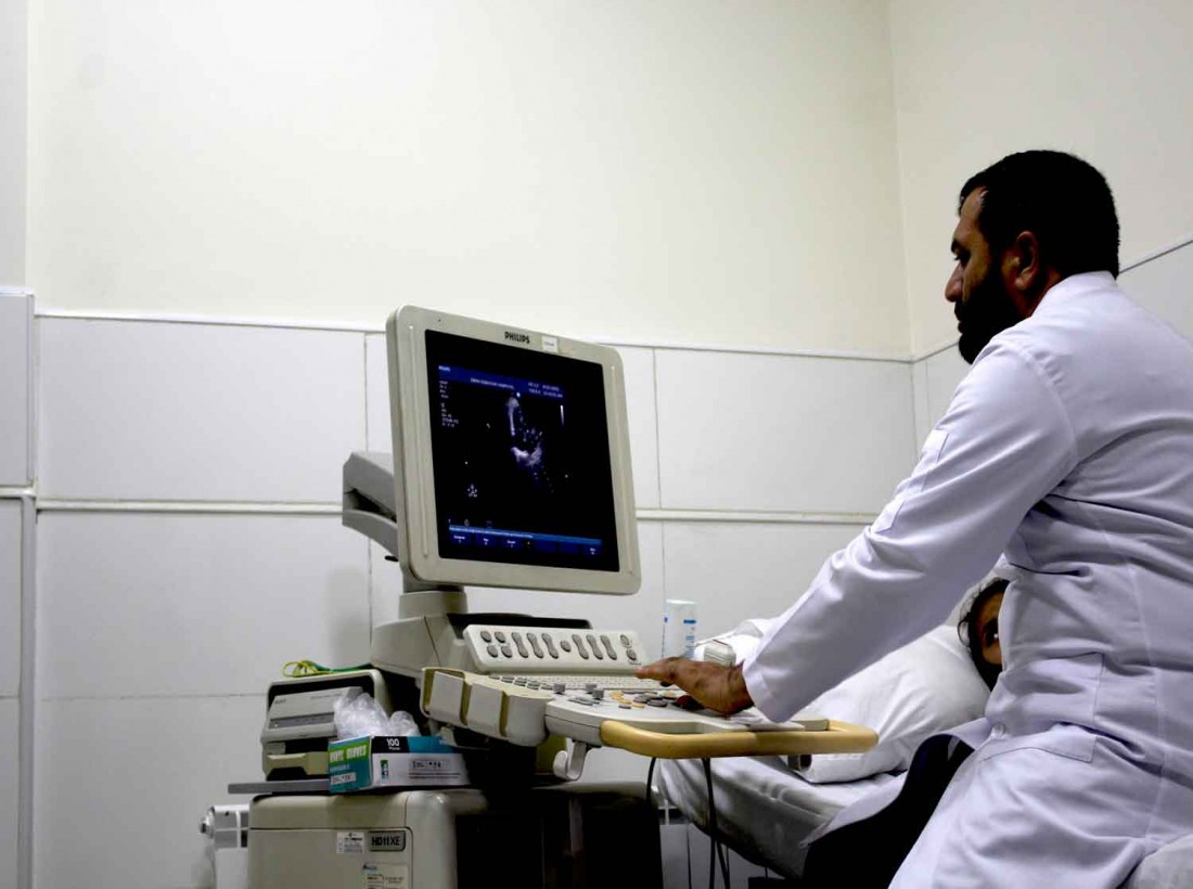
An echocardiogram uses sound waves to produce images of your heart. This common test allows your doctor to see your heart beating and pumping blood. Your doctor can use the images from an echocardiogram to identify heart disease.
Depending on what information your doctor needs, you may have one of several types of echocardiograms. Each type of echocardiogram involves few, if any, risks.
Your doctor may suggest an echocardiogram to:
The type of echocardiogram you have depends on the information your doctor needs.
In this standard type of echocardiogram:
If your lungs or ribs block the view, you may need a small amount of an enhancing agent injected through an intravenous (IV) line. The enhancing agent, which is generally safe and well tolerated, will make your heart's structures show up more clearly on a monitor.
If your doctor wants more-detailed images or it's difficult to get a clear picture of your heart with a standard echocardiogram, your doctor may recommend a transesophageal echocardiogram.
In this procedure:
Sound waves change pitch when they bounce off blood cells moving through your heart and blood vessels. These changes (Doppler signals) can help your doctor measure the speed and direction of the blood flow in your heart.
Doppler techniques are generally used in transthoracic and transesophageal echocardiograms. Doppler techniques can also be used to check blood flow problems and blood pressure in the arteries of your heart — which traditional ultrasound might not detect.
The blood flow shown on the monitor is colorized to help your doctor pinpoint any problems.
Zwan Medical Complex's Covid-19 PCR test accepted across all the flight agencies in Afghanistan. What is a PCR test? PCR… ...
Read More
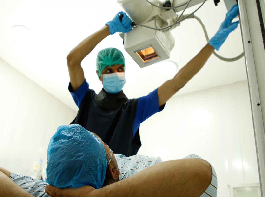
Who are we? An x-ray is a widely used test which creates an image of the inside of the body, much like a photograph. It… ...
Read More
An echocardiogram uses sound waves to produce images of your heart. This common test allows your doctor to see your heart… ...
Read More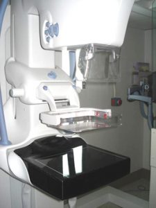
Mammography Breast cancer is a fierce enemy. It remains the most frequently diagnosed non-skin cancer in women, with more… ...
Read More

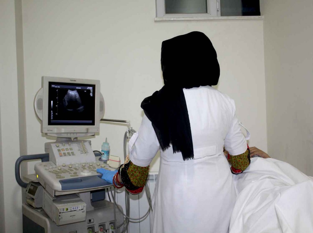
What is ultrasound?An ultrasound scan is a painless test that uses sound waves to make images of organs and structures inside… ...
Read More
Laboratory services are provided in a well-equipped, highly automated and professional center. Normal and specialized diagnostic… ...
Read More